Trichoscopy & Digital Analysis
Clinical methods for diagnosing hair loss using imaging analysis.
Principles of Trichoscopy
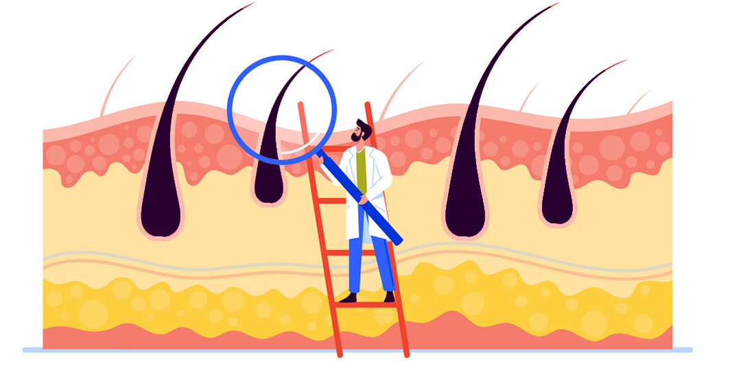
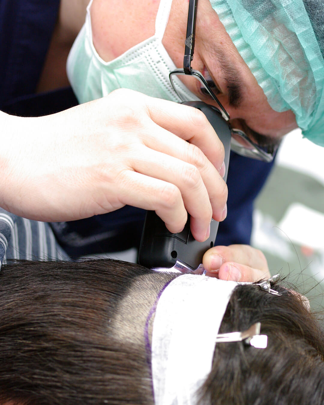
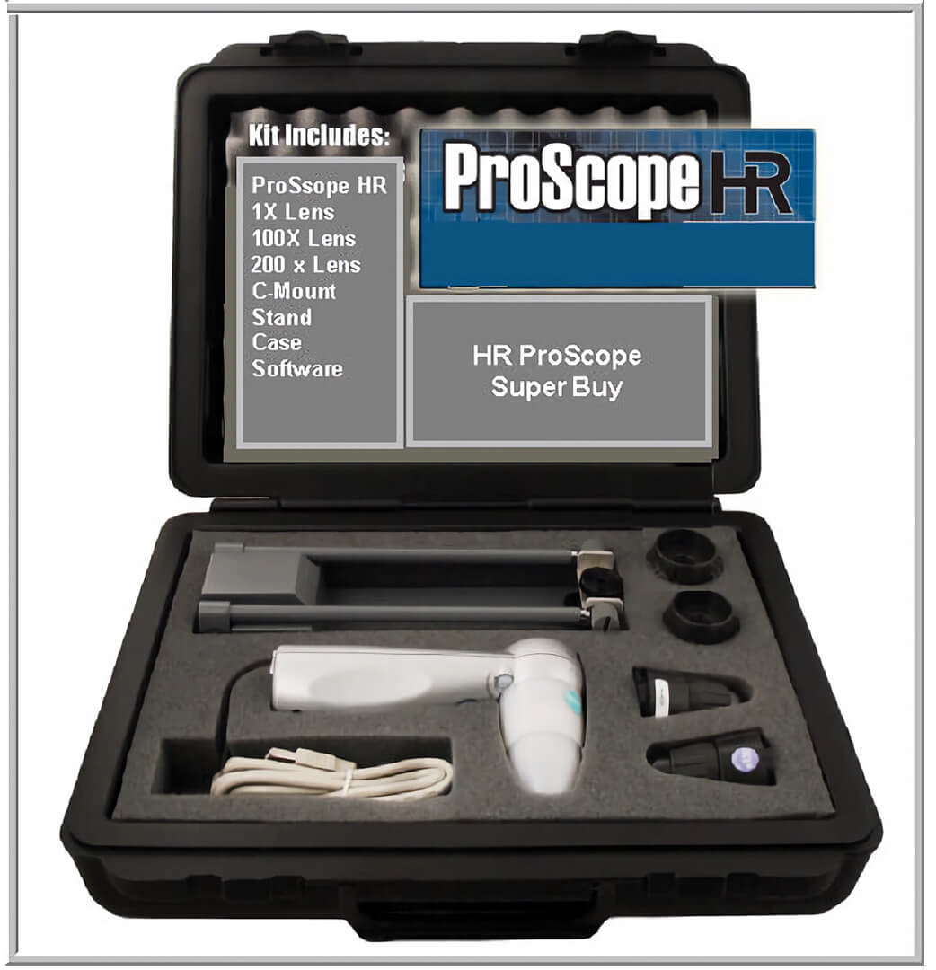
Trichoscopy, also known as dermoscopy of the hair and scalp, is a non-invasive diagnostic method using polarized or non-polarized light magnification. It visualizes hair shafts, follicular openings, and vascular patterns.
Clinical Utility
- Differential diagnosis: Distinguishing between scarring and non-scarring alopecias.
- Activity assessment: Identifying signs of inflammation that may affect treatment planning.
- Treatment monitoring: Documenting changes in hair density and shaft diameter.
- Biopsy guidance: Selecting optimal sites for tissue sampling.
This non-invasive visualization is a standard component of hair loss evaluation.
Scarring vs. Non-Scarring Alopecia
This distinction helps determine treatment pathways.
Non-Scarring Alopecia
- Follicular openings are visible and intact.
- Hairs thin but follicles remain.
- Examples: Androgenetic alopecia, alopecia areata, telogen effluvium.
Scarring Alopecia
- Fibrosed or destroyed follicular openings.
- Permanent absence of follicular pores.
- Examples: Lichen planopilaris, frontal fibrosing alopecia.
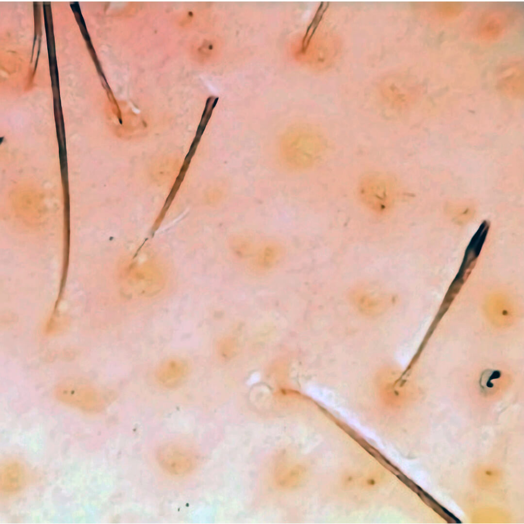
Trichoscopic Patterns by Condition
Androgenetic Alopecia
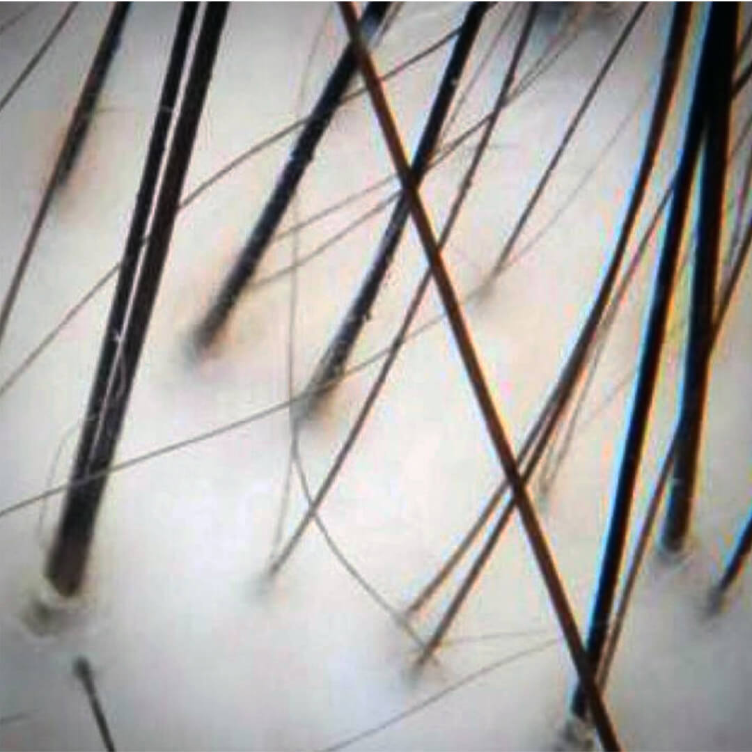
- Hair diameter diversity greater than 20%
- Peripilar signs
- Yellow dots
- Vellus hairs
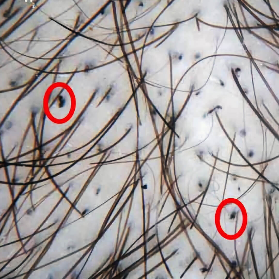
Alopecia Areata
- Yellow dots
- Exclamation mark hairs
- Black dots (broken hairs)
- Cadaverized hairs
Scarring Alopecias (LPP, FFA)
- White dots (fibrosed follicular openings)
- Perifollicular scaling
- Erythema
- Loss of follicular openings
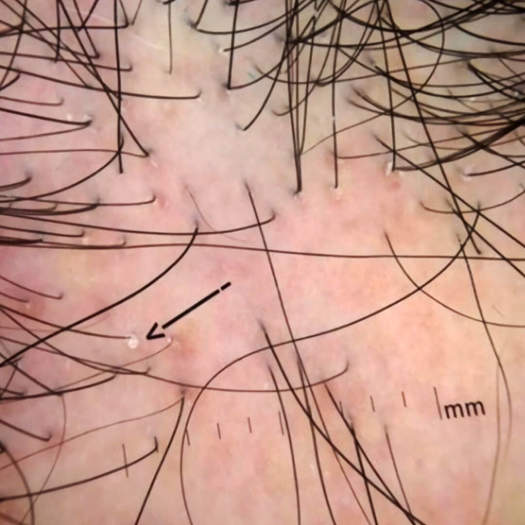
Trichotillomania
- Broken hairs of varying lengths
- Flame hairs
- Coiled or tulip hairs
- V-sign (split ends)
Key Clinical Points
- Trichoscopy is a first-line, non-invasive diagnostic tool for hair loss evaluation.
- Androgenetic alopecia shows hair diameter diversity greater than 20% and increased vellus hairs.
- Alopecia areata presents with yellow dots and exclamation mark hairs.
- Scarring alopecias show white dots, loss of follicular openings, and perifollicular scaling.